📞 88558 10010 Silver Leaf Clinic 📞 +91 20 6768 9704 Landline 📞 84118 08284 Dr. Gore / WhatsApp
Silver Leaf Clinic · Hadapsar, Pune · Colorectal & GI Cancer Centre
Home Procedures ICG Fluorescence
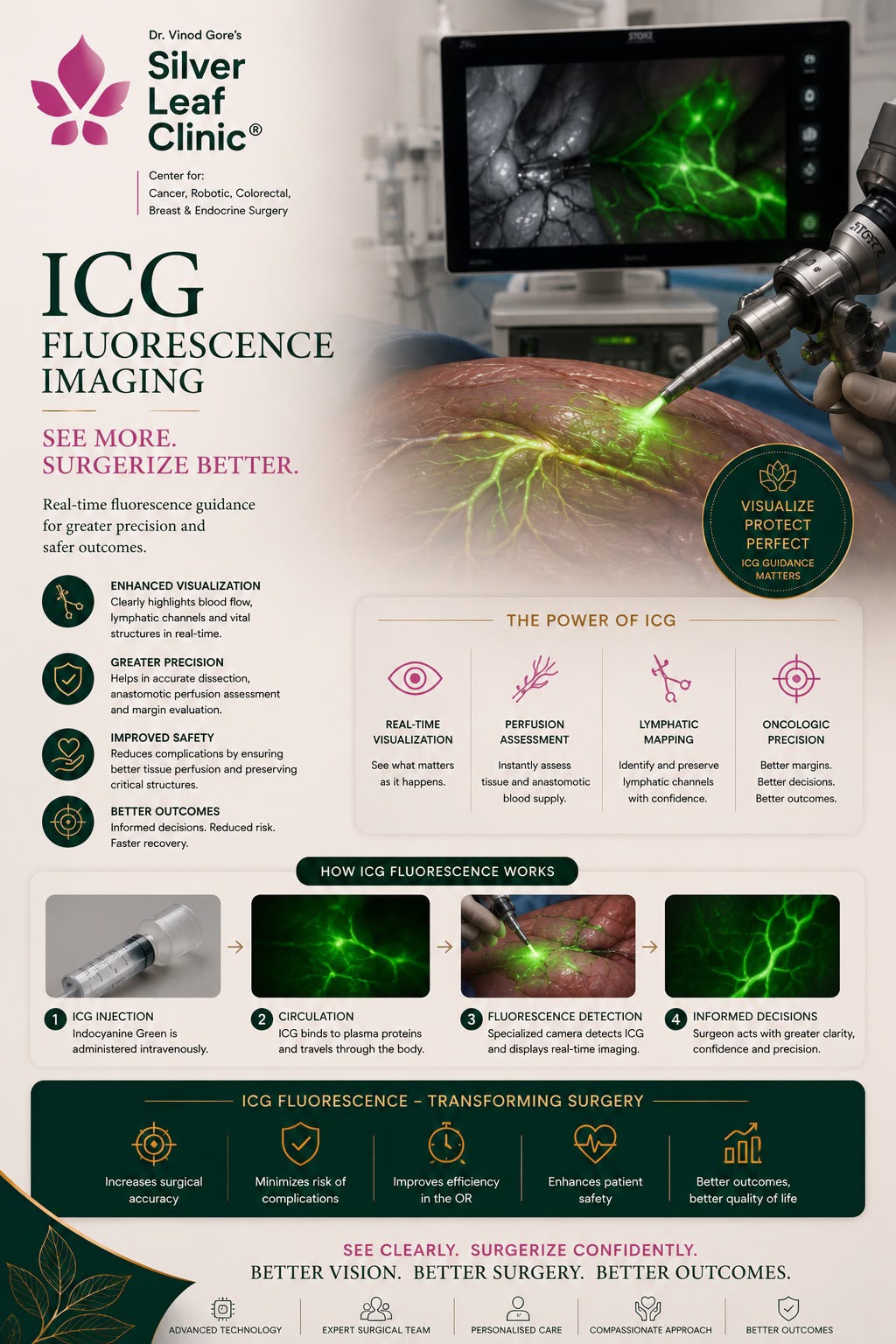
Indocyanine Green · Near-Infrared Imaging

ICG Fluorescence

Seeing the Unseen in GI & Colorectal Surgery · Pune

ICG fluorescence lets the surgeon see things the naked eye cannot — whether bowel is getting a good blood supply, where a tumour ends, which lymph node drains it, and where delicate structures lie. A harmless green dye glows under near-infrared light, turning invisible anatomy into a live, glowing map during surgery. Dr. Vinod T. Gore uses ICG routinely — with the Stryker SPY system in open and laparoscopic surgery, and the built-in Firefly camera on the da Vinci Xi robot — to make cancer surgery safer and more precise.

Real-Time Imaging Stryker SPY da Vinci Firefly Open · Lap · Robotic
Rectal Cancer Surgery - Dr. Vinod T. Gore
The Basics

What is ICG — and what is fluorescence?

ICG (indocyanine green) is a safe, water-soluble dye that has been used in medicine for decades. Given as a small injection into a vein — or sometimes around a tumour — it travels through the bloodstream and lymphatic channels, and is cleared quickly and harmlessly by the liver. Allergic reactions are very rare.

Fluorescence means the dye glows when a particular light is shone on it. ICG absorbs near-infrared (NIR) light — a wavelength invisible to our eyes — and re-emits it as a bright signal that a special camera detects and displays, usually as a vivid green overlay on the surgeon's screen.

How it works in surgery: the surgeon injects ICG, switches the camera to near-infrared mode, and within seconds the structures carrying the dye light up. Well-perfused bowel glows brightly; poorly perfused tissue stays dark. Lymph nodes draining a tumour light up. A liver segment fed by one vessel glows while neighbours stay dark. It is real-time, repeatable, and adds only minutes to the operation.

In short, ICG turns the surgeon's judgement from "this looks healthy" into "I can see it is healthy" — objective information, exactly when the decision is being made.

Why it matters for your safety
Many of the most serious complications in GI and colorectal surgery come from a join (anastomosis) that did not have a good enough blood supply to heal. ICG lets the surgeon check perfusion before committing to the join — and change the plan if needed. It is one of the simplest, safest ways to reduce leaks and make cancer surgery more precise.
The Technology

Two systems — every approach covered

ICG fluorescence is not limited to one type of surgery. Dr. Gore uses it across open, laparoscopic and robotic operations, with the right system for each.

Stryker SPY system
A near-infrared imaging platform used in open and laparoscopic surgery. A handheld or laparoscopic NIR camera visualises ICG perfusion and structures on screen — bringing fluorescence guidance to conventional operations, not just robotic ones.
da Vinci Xi · Firefly
The da Vinci Xi robot has ICG fluorescence (Firefly) built in. With one click the surgeon toggles between normal and near-infrared 3-D vision at the console — seamless, magnified, fluorescence-guided robotic surgery.
Open · Lap · Robotic
Because both systems are available, the benefits of ICG are not restricted to expensive robotic cases — patients having open or keyhole surgery get the same fluorescence-guided safety.
Indications

Where ICG is used in GI & colorectal surgery

ICG has become a versatile tool across the whole field — from checking blood supply, to mapping lymph nodes, to guiding liver resection. These are its main uses.

Rectal Cancer Surgery - Dr. Vinod T. Gore
ICG fluorescence — indications in GI & colorectal surgery
UseWhat ICG showsBenefit to the patient
Bowel & anastomotic perfusionWhich segments of bowel have a strong blood supply and which are poorly perfusedReduces the risk of an anastomotic leak — the most serious complication after bowel surgery
Choosing the resection lineThe exact point where blood supply is healthy, before the bowel is dividedThe join is made in well-perfused tissue, helping it heal soundly
Sentinel lymph node mappingThe first lymph node(s) draining a tumour, glowing along lymphatic channelsGuides accurate nodal assessment and more tailored lymph node surgery
Thoracic duct identification
(esophagectomy)
The thoracic duct lit up during oesophageal surgeryAvoids injury to the duct, preventing a chyle leak — a difficult post-op problem
Tumour & metastasis detectionTumour deposits and small surface metastases that retain dyeFinds disease the eye and scans can miss, making clearance more complete
Liver tumour marginsThe edge of a liver tumour against healthy liverHelps remove the tumour with a clear margin while sparing healthy liver
Liver segment delineationThe exact territory of a liver segment fed by one vessel (it glows; neighbours stay dark)Enables precise anatomical resection and parenchyma-sparing surgery
Ureter & biliary mappingThe ureters or bile ducts highlighted (with specific ICG techniques)Protects these delicate structures from accidental injury

One dye, many answers. The same simple injection can, in different settings, check blood supply, map lymph nodes, find tumour and guide liver resection — which is why ICG has quietly become one of the most useful tools in modern cancer surgery.

In Practice

How ICG fits into an operation

Using ICG is quick and woven naturally into the flow of surgery — here is a typical example during a colorectal resection.

Before dividing
Check perfusion & pick the line
ICG is injected and the bowel is viewed in near-infrared. The surgeon sees exactly where blood supply is healthy and chooses the resection line there.
During
Map nodes or structures
Where relevant, sentinel nodes are mapped, or delicate structures (thoracic duct, ureters, bile ducts) are highlighted to avoid injury.
Before the join
Confirm both ends glow
Before creating the anastomosis, both ends are re-checked in fluorescence — only well-perfused tissue is joined, lowering leak risk.
Liver cases
Define margins & segments
In liver surgery, ICG marks the tumour edge and lights up the segment to be removed, guiding a precise, parenchyma-sparing resection.
Patient Questions

ICG fluorescence — answered plainly

Yes. ICG has been used safely in medicine for decades. It is given in a tiny dose, cleared quickly by the liver, and serious allergic reactions are very rare. It carries no radiation. The main caution is for people with a known iodine allergy, which we always check beforehand.
No — it adds only a few minutes and makes surgery safer, not riskier. The extra information it gives, especially about blood supply before a join, helps prevent complications like leaks.
Both. With the Stryker SPY system, ICG fluorescence is available in open and laparoscopic surgery, and it is built into the da Vinci Xi robot as Firefly. So the benefit is not limited to robotic cases — patients having any approach can gain from it.
A bowel join only heals well if both ends have a good blood supply. ICG lets the surgeon see perfusion directly and choose to join only healthy, well-perfused tissue — and to change the plan if an area looks poorly supplied. This is one of its most valuable uses.
In two ways: it outlines the edge of a liver tumour so it can be removed with a clear margin, and it lights up the exact liver segment supplied by one vessel — so the surgeon can perform a precise anatomical resection that removes the tumour while sparing as much healthy liver as possible.

This page is general information, not personal medical advice. The use of ICG is decided by the surgeon for each operation. Please discuss your specific surgery at your consultation.

Silver Leaf Clinic · Hadapsar, Pune

Surgery guided by what we can see

Dr. Gore uses ICG fluorescence routinely to make GI and colorectal cancer surgery safer and more precise — across open, laparoscopic and robotic approaches. To discuss your surgery, please bring all CT/MRI/PET scans, biopsy reports and previous operative notes — ideally on CD or shared via WhatsApp in advance.

📍 Clinic Address
511, City Centre, Solapur Road, Opp. Vaibhav Theatre, Hadapsar, Pune 411028
📍 Open in Google Maps →
Monday – Saturday10:00 AM – 6:00 PM
SundayBy prior appointment
Surgical Hospital
Sahyadri Manipal Hospital, Pune · Robotic Cancer Surgery Centre of Excellence